13929258449

admin@satnano.com

Nanotechnology exchange

Home

Nanotechnology exchange

How to Prepare of Samples for Nanoparticle Transmission Electron Microscopy (TEM)

July 1,2022.
Transmission Electron Microscope (TEM)

Transmission electron microscopy uses an electron beam that penetrates a sample to image, which requires the sample being observed to be "transparent" to the incident electron beam.

Transmission electron microscopy is widely used in materials science and biology. Since electrons are easily scattered or absorbed by objects, the penetration is low, and the density and thickness of the sample will affect the final imaging quality. Thinner ultrathin sections must be prepared, usually 50-100 nm. Therefore, the sample for observation by transmission electron microscope needs to be processed very thinly. Commonly used methods are: ultra-thin sectioning, frozen ultra-thin sectioning, freeze-etching, freeze-fracture and so on.

For powder samples, the samples can be prepared by ultrasonic dispersion.

For liquid samples or disperse samples, they can be dropped directly on the copper mesh.

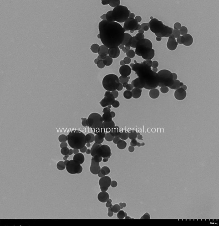
Picture:Silicon Nanoparticles Transmission Electron Microscope TEM


At lower magnifications, the contrast in TEM imaging is mainly due to the different absorption of electrons due to the different thickness and composition of the material.

When the magnification is high, the complex fluctuation will cause the difference in the brightness of the image, so professional knowledge is needed to analyze the obtained image. By using different modes of TEM, a sample can be imaged by its chemical properties, crystallographic orientation, electronic structure, electronic phase shift caused by the sample, and generally by electron absorption.

Leave a message Please click here for inquiry
Provide the right solution according to your needs,offer efficient service,leave a message for the product information and requirements you need,customize now!